
INTRODUCTION
The window of implantation (WOI) paradigm has shaped reproductive medicine for over three decades, positing that successful human embryo implantation occurs during a narrow, hormonally regulated period of endometrial receptivity. Authoritative sources have defined this window spanning various days of a 28-day cycle: days 20–24,1 days 22–24,2 or beginning on cycle day 19–20 and lasting 4–5 days.3 Whatever the precise range, the clinical implication is unambiguous: embryo transfer outside this period is considered to be incompatible with pregnancy. This theory has driven the development of endometrial receptivity assays, personalized embryo transfer protocols, and WOI-based interventions that form a cornerstone of modern assisted reproductive technology (ART) practice.4,5
A fundamental biological occurrence contradicts this paradigm. Despite decades of WOI-focused ART optimization, even chromosomally normal embryos fail to implant in approximately 35–50% of transfers, a gap that has recently been described as “the black box of implantation”.6 At the same time, extrauterine pregnancies occur in approximately 1–2% of conceptions,7,8 successfully establishing and developing in anatomical sites completely devoid of endometrial preparation. Locations include the fallopian tubes (93–97% of ectopic cases), ovaries, omentum, peritoneum, abdominal organs, and even neoplastic endometrial tissue.9–11 More striking still, rare cases have progressed to advanced gestational ages, including ovarian pregnancies reaching 34 weeks12 and full term,13 tubal pregnancies advancing to the third trimester,14 and abdominal pregnancies that delivered live neonates at 33 weeks,15 40+4 weeks,16 and 41 weeks.17
This presents a critical paradox with immediate clinical implications: if implantation truly required the precise molecular and temporal conditions of the endometrial WOI, how can extrauterine pregnancies occur at all, let alone progress to viability? The simultaneous phenomena of successful ectopic implantation and persistent ART failures suggest that either fundamental gaps exist in our understanding of implantation requirements or that the WOI offers optimal rather than exclusively required conditions for pregnancy establishment.
The central observation at the core of this critical review is straightforward. The conditions sufficient for embryonic attachment must be distinguished from the conditions required to sustain a pregnancy to term. The fallopian tube constitutively expresses integrin receptors (α1β1, α4β1, and αvβ3) and their ligand osteopontin, with no evidence of cyclical changes during the mid-luteal phase.18 This constitutive molecular expression demonstrates that the machinery that permits trophoblast attachment is not confined to a narrow temporal window in a single tissue type. The endometrium is exceptional not because it alone enables attachment, but because it additionally provides the decidualized stromal architecture, organized spiral artery remodeling, and immune-regulatory environment required to carry a pregnancy to term.1,19 The near-universal failure of extrauterine pregnancies to reach viability, despite successful initial implantation, powerfully illustrates that it is this sustained developmental support, not the act of attachment itself, that the uterine environment uniquely provides.
What, then, are the minimum conditions any tissue must satisfy for trophoblastic implantation to occur? The pattern emerging from the extrauterine pregnancy literature suggests two: adequate vascularity sufficient to perfuse a developing trophoblast, and exposure to the systemic mid-luteal hormonal milieu, in particular the elevated circulating progesterone produced by a functioning corpus luteum at the time of attachment.20 These two conditions are not unique to the endometrium; they are properties of the systemic state of early pregnancy and of any well-perfused tissue exposed to it. The endometrium uniquely provides the third, separable element: the decidualized matrix required to sustain pregnancy to term.
If this reframing is correct, the WOI may not function as the precise temporal gate that endometrial receptivity assays purport to measure. The largest randomized trial (RCT) of endometrial receptivity array (ERA)-guided personalized embryo transfer found no improvement in live birth rates compared with standard timing,21 and independent reanalysis concluded that the ERA failed to identify the WOI as purported.22 Multiple systematic reviews corroborate this finding.23–25 With repeated euploid transfers, cumulative live birth rates reach 98.1%,26 which further suggests that most implantation failures are stochastic rather than deterministic. In addition, the Lugano Workshop consensus concluded that true recurrent implantation failure (RIF), defined as the failure of three consecutive euploid single embryo transfers, affects fewer than 5% of couples.27 If embryos can attach to the fallopian tube epithelium, ovarian stroma, omentum, and peritoneal surfaces – all tissues with no temporal receptivity programming whatsoever – the insistence on pinpointing a precise endometrial window warrants critical reexamination.
This review tests three competing hypotheses about implantation biology. The conventional model holds that implantation requires specific endometrial preparation during a narrow temporal window. An embryo-centric model proposes that implantation is primarily an embryo-driven process that can occur across diverse tissue sites when minimal supportive conditions are met. A hybrid model, which the evidence on extrauterine pregnancy most strongly supports, holds that endometrial preparation optimizes the probability of implantation and is essential for sustained pregnancy, but is not an absolute requirement for attachment itself. By systematically examining these “natural experiments,” this review evaluates whether evidence from extrauterine pregnancies supports a biologically separable model of implantation, one in which permissive trophoblastic attachment and sustained gestational support are distinct functions with different tissue requirements. We then consider the implications of this framework for current ART practice.
CRITICAL SYNTHESIS OF EVIDENCE FROM EXTRAUTERINE PREGNANCIES
Frequency and Anatomical Distribution
As discussed in the Introduction, ectopic pregnancies occur in approximately 1–2% of all pregnancies, posing a substantial global burden. The Global Burden of Disease 2021 analysis estimated that approximately 8.38 million ectopic pregnancy cases occurred worldwide in 2021.28 Ectopic pregnancies remain the leading cause of first-trimester pregnancy-related death, responsible for up to 6% of maternal mortality during early gestation29; non-tubal ectopic pregnancies carry a disproportionately higher fatality rate than tubal pregnancies.30
A natural objection to the central thesis of this review is the following: if implantation is so permissive of non-endometrial sites, why are ectopic pregnancies not far more common? The answer lies primarily in anatomy and embryo transport rather than in tissue-specific restrictions on receptivity. Ectopic pregnancies result from impaired embryo transport, including tubal inflammation, scarring, altered motility, and ciliary dysfunction, rather than from WOI-related mechanisms.7,29 The fallopian tube functions as an active transport system in which ciliated epithelium and coordinated peristalsis deliver the blastocyst to the uterine cavity over several days. When this transport functions normally, the developing embryo never encounters non-endometrial tissue. The classic risk factors for ectopic pregnancy, including pelvic inflammatory disease, prior tubal surgery, and altered tubal motility, are precisely those that compromise transport.7 Notably, ectopic pregnancies still occur following in vitro fertilization (IVF) embryo transfer (ET), when embryos are deposited directly into the uterine cavity. Moreover, tubal pregnancies following IVF-ET demonstrate markedly stronger E-cadherin immunostaining in chorionic villi trophoblasts than do spontaneous tubal pregnancies, suggesting that embryos actively participate in establishing adhesion in non-endometrial sites.31 Heterotopic pregnancies, in which intrauterine and ectopic implantation occur simultaneously within the same cycle, demonstrate that the same systemic hormonal milieu simultaneously supports successful trophoblastic attachment in two anatomically and histologically distinct tissues.32 These observations are difficult to reconcile with a model in which the endometrium possesses a uniquely narrow receptivity window for embryonic attachment. Ectopic pregnancy is therefore best understood not as a receptivity exception but as a transport failure that exposes the trophoblast’s inherent permissiveness (its capacity for attachment across tissue types), normally concealed by the efficient delivery of the embryo to its default uterine destination. Table 1 below compares implantation environments and clinical outcomes.
Anatomical Sites and Survival Patterns
The literature indicates that extrauterine implantation occurs at remarkably diverse anatomical sites, each offering distinct insights into the minimal requirements for embryonic attachment.
As noted above, fallopian tube pregnancies constitute 93–97% of ectopic cases. The limited vascular supply of the fallopian tube cannot sustain trophoblastic growth indefinitely, and most tubal pregnancies are diagnosed between 6 and 10 weeks of gestation, either by clinical detection or by rupture.7 However, the initial success of tubal implantation is itself informative. As discussed above, the fallopian tube epithelium expresses integrin receptors and osteopontin constitutively, with minimal cyclical regulation. This constitutive molecular expression, in contrast to the cyclical upregulation that defines the endometrial WOI, demonstrates that the molecular machinery that permits trophoblast attachment can exist independently of temporal regulation. Notably, rare tubal pregnancies have advanced well beyond the first trimester, with documented cases reaching the third trimester.14,34
As quoted above by Long et al., ovarian pregnancies account for 1–6% of all ectopic pregnancies, providing some of the most compelling evidence for implantation flexibility. Previously cited cases include a nearly viable 34-week pregnancy and full-term ovarian pregnancies with live neonates. Uniquely among adult human tissues, the ovary exhibits cyclical physiological angiogenesis driven by vascular endothelial growth factor (VEGF) and fibroblast growth factor 2 (FGF2),35 providing a particularly favorable microenvironment. This may explain the over-representation of ovarian cases among extrauterine pregnancies reaching advanced gestational ages.
Abdominal pregnancies account for 0.3–1.4% of all ectopic pregnancies30 and encompass implantation on the peritoneum, omentum, bowel serosa, and solid organ surfaces. Documented cases, cited above, include live neonates delivered at or near term, though such outcomes carry extreme maternal morbidity and mortality.36,37 Advanced abdominal pregnancies typically develop secondary placental attachments to well-vascularized structures such as the bowel mesentery or broad ligament, effectively co-opting existing vascular networks to sustain fetal growth.10 Omental pregnancies are among the rarest subtypes, with most reported cases presenting as emergency ruptures in the first trimester.38 The omentum bears no structural resemblance to the endometrium yet possesses unique immune-regulatory properties through milky spots, organized aggregates of macrophages, B cells, and T cells.39
Other rare implantation sites further illustrate trophoblast adaptability. Hepatic ectopic pregnancies have been documented, with an estimated incidence of approximately 1 in 15,000 pregnancies.40 Splenic ectopic pregnancy, though exceedingly rare, has also been reported.9,41 Perhaps most compelling is the documentation of embryonic implantation directly within endometrial carcinoma tissue, discovered during surgery for a presumed tubal pregnancy,11 demonstrating that trophoblast attachment can proceed even in neoplastic tissue.
Table 2 outlines extrauterine implantation sites by frequency and progression.
Mechanisms Enabling Implantation in Non-Endometrial Tissue
Successful implantation, whether intrauterine or ectopic, depends on coordinated immune tolerance, adequate vascularization, and structural support for embryonic development. In the uterus, decidual natural killer (dNK) cells constitute 50–70% of decidual lymphocytes during the first trimester.44 These dNK cells are poorly cytolytic, instead releasing cytokines and chemokines that induce trophoblast invasion, tissue remodeling, and placentation.44 Regulatory T cells (Tregs) and macrophages further contribute to fetomaternal tolerance, with the fetal placenta itself, mainly through trophoblast cells, inducing homeostatic M2 macrophages and functional Tregs via placental-derived macrophage colony-stimulating factor (M-CSF) and interleukin-10 (IL-10).45 Critically, this placental capacity to generate immune tolerance is an autonomous property of trophoblast cells rather than a function of the uterine environment per se, as demonstrated by in vitro studies using placental tissue.45 The survival of ectopic pregnancies is consistent with this autonomous immunomodulatory capacity: in tubal pregnancies, reduced apoptosis of extravillous trophoblast cells compared with intrauterine implantation suggests that the distinct immunological microenvironment at ectopic sites fails to limit trophoblast invasion, which may paradoxically facilitate initial implantation success.46 The fallopian tube notably lacks NK cells in its mucosa, and tubal ectopic pregnancy is characterized by elevated levels of pro-inflammatory cytokines (IL-6, IL-8) produced by macrophages, which paradoxically create a pro-implantation rather than a tissue-rejection environment.47
Vascular development is a critical determinant of ectopic pregnancy survival. While most ectopic sites lack the organized spiral artery remodeling characteristic of intrauterine pregnancy,48 at tubal implantation sites, VEGF and its receptors, kinase insert domain receptor (KDR) and fms-like tyrosine kinase 1 (Flt-1), are markedly upregulated, with serum VEGF correlating with the depth of trophoblastic invasion.49,50 Adhesion molecule profiles are similarly remodeled at ectopic sites: E-cadherin expression is significantly reduced at tubal implantation sites compared with non-implantation segments, while β1 integrin and phosphorylated focal adhesion kinase are significantly upregulated51; this mirrors the epithelial-mesenchymal transition that characterizes invasive trophoblast differentiation in intrauterine pregnancy.52 Mucin 1 (MUC1), a glycoprotein barrier to implantation, is significantly decreased in fallopian tubes bearing ectopic pregnancies, while pro-implantation factors FGF2 and heparin-binding epidermal growth factor-like growth factor (HB-EGF) are upregulated at the tubal implantation site.53
Trophoblast invasion shows striking parallels with cancer cell metastasis: both employ matrix metalloproteinases to degrade tissue barriers, stimulate angiogenesis via VEGF production, and evade immune rejection through immunosuppressive factor secretion.54–56 This shared invasive toolkit, evolved to penetrate the decidualized endometrium and remodel maternal spiral arteries,57 retains its capacity wherever the trophoblast lands. Ectopic pregnancy may thus be understood not as a failure of the implantation program but as that program operating in the wrong anatomical location, an inevitable trade-off of the highly invasive hemochorial placentation strategy that humans share with other great apes.
Table 3 compares the molecular and cellular qualities of uterine versus ectopic implantation sites.
The Common Denominator: Vascularity and Systemic Mid-Luteal Progesterone
A striking pattern emerges when the extrauterine implantation sites described above are examined collectively. Despite their profound histological differences, including ciliated columnar epithelium in the fallopian tube, mesothelial covering over a vascular stroma in the ovary, adipose tissue with milky spots in the omentum, simple mesothelium on the peritoneum and bowel serosa, and even neoplastic glandular epithelium in the case of implantation within endometrial carcinoma, these tissues share two functional features that the endometrium also possesses: sufficient vascularization to support trophoblast growth and appropriate systemic hormonal exposure. We propose that these two features, rather than the specific molecular signature of decidualization, constitute the minimal sufficient conditions for trophoblastic attachment.
Vascular sufficiency emerges as the principal determinant of how far an extrauterine pregnancy can progress. At tubal implantation sites, VEGF and its receptors are markedly upregulated, and circulating VEGF correlates with the depth of trophoblastic invasion into the tubal wall.49,50 The ovary’s cyclical physiological angiogenesis35 provides a particularly favorable microenvironment, which helps explain why ovarian pregnancies are over-represented among cases that reach advanced gestational ages.12,13,62
As stated above in the Anatomical Sites and Survival Patterns section, the omentum carries a dense vascular network organized around milky spots; abdominal pregnancies that reach viability typically establish secondary placental attachments to highly vascularized peritoneal structures rather than poorly perfused surfaces.15,17 The histological analysis of the 34-week ovarian pregnancy reported by Meseci and colleagues showed extensive vascular networks and robust angiogenesis at the implantation site, leading those authors to conclude that vascular sufficiency, rather than tissue identity, is the primary constraint on extrauterine pregnancy survival.12 The corollary observation strengthens this interpretation, as noted previously by Schreiber and Sonalkar: extrauterine pregnancies fail early most often when implanted at sites of poor vascular access.
The hormonal dimension is equally important and, until now, has yet to receive the attention it warrants in the implantation literature. Like intrauterine pregnancies, ectopic pregnancies occur in the cycle following ovulation and require a normally functioning corpus luteum; hCG levels rise initially with normal doubling times, and serum progesterone reaches its physiological mid-luteal peak at the moment of trophoblast attachment.20 The trophoblast attaches in a tissue that, regardless of its identity, is bathed in a systemic hormonal milieu rich in progesterone, estrogens, and corpus-luteum-derived peptides. Decidualization of the endometrium is the local expression of this systemic milieu acting on a primed stromal compartment. Still, the systemic milieu itself reaches every vascularized tissue in the body, including the fallopian tube, ovary, omentum, and peritoneum. This explains a paradox the conventional WOI model cannot easily accommodate: how trophoblastic attachment succeeds reliably in tissues that never undergo decidualization. The local molecular outputs of decidualization, including secretory transformation, immunomodulatory cytokines, and changes in vascular permeability, may be partially reproduced at extrauterine sites by the embryo itself, acting on tissues already preconditioned by systemic mid-luteal progesterone.
Taken together, these observations support a reframing of the minimal conditions for implantation. The endometrial WOI is best understood not as a tissue-specific molecular gate, but as the local function in one particular tissue of two more fundamental requirements that multiple non-endometrial tissues appear capable of satisfying. These are the vascular supply and systemic hormonal milieu, as established above. The endometrium is uniquely qualified to provide a third element, as explained in the Introduction, the decidual support required to carry a pregnancy to term; however, this is a separate requirement from whether attachment can occur. The clinical record of extrauterine pregnancies indicates that it can.
A Two-Stage Model of Human Implantation
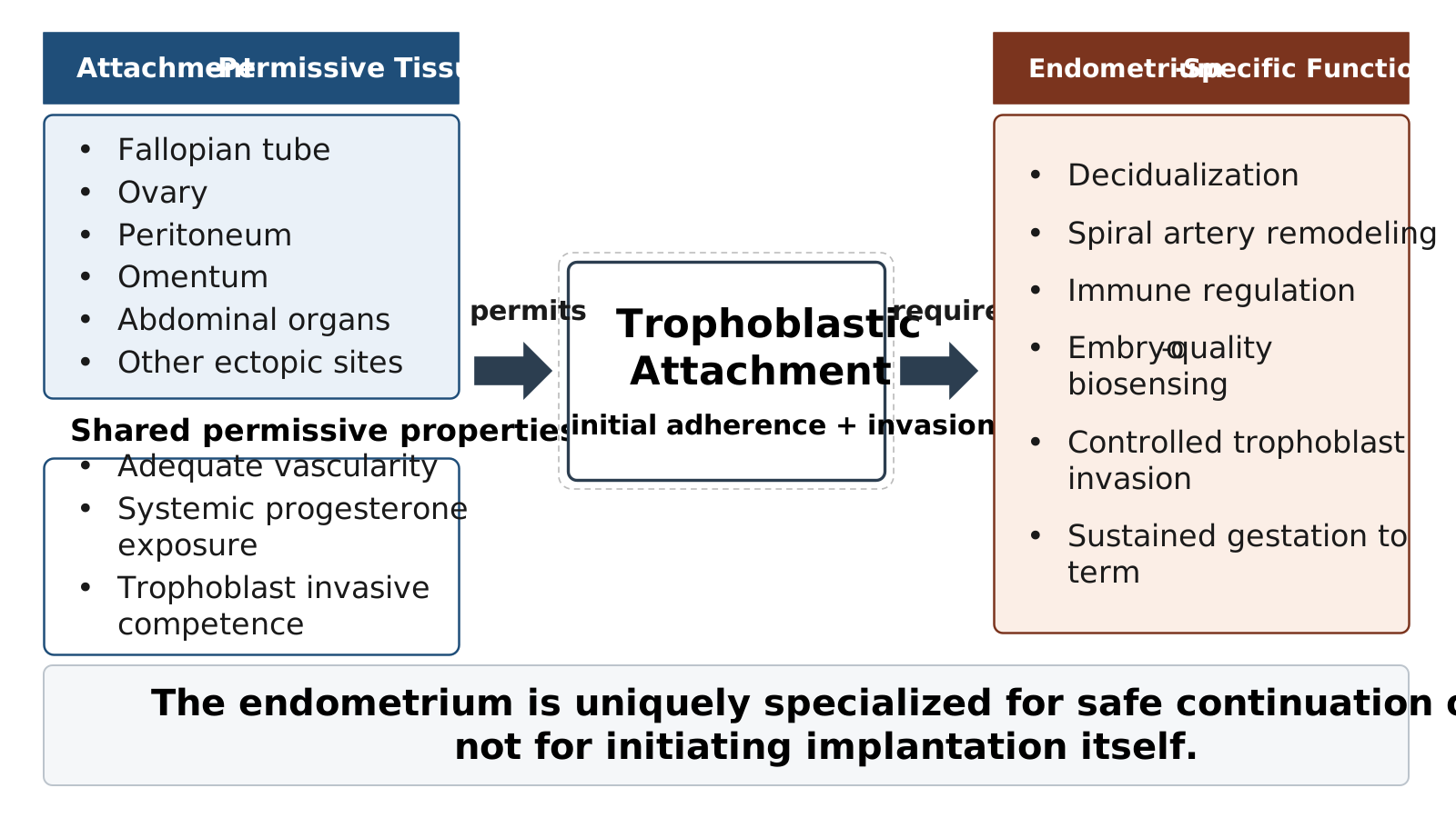
The evidence reviewed throughout this manuscript supports a conceptual reframing of human implantation as a biologically separable two-stage process rather than a single unified event. Current WOI models implicitly treat embryonic attachment and successful continuation of pregnancy as manifestations of the same requirement: a precisely timed state of endometrial receptivity. The literature on ectopic pregnancy suggests instead that these represent fundamentally distinct biological processes with different requirements.
The first stage – trophoblastic attachment – is governed by the two permissive conditions identified above: vascular sufficiency and the hormonal milieu. As demonstrated by the diversity of extrauterine implantation sites, these conditions are not unique to the endometrium. The trophoblast itself supplies much of the invasive, angiogenic, and immunomodulatory machinery required for initial attachment.
The second stage – sustained gestational support – is highly specialized to the decidualized endometrium, which, as previously established, uniquely drives spiral artery remodeling, decidual stromal architecture, controlled immune tolerance, regulation of trophoblast invasion, and embryo-quality biosensing. Because these functions are not performed at extrauterine sites, ectopic pregnancies do not progress safely. This also distinguishes the endometrium not as the exclusive initiator of implantation but as the uniquely evolved organ capable of sustaining pregnancy.
Within this framework, the WOI is the endometrium’s optimized version of biological conditions that permit attachment, rather than a prerequisite for them. Ectopic pregnancies confirm that biological permissiveness for attachment is broader than the specialized conditions required for safe continuation of pregnancy.
Table 4 presents empirical evidence supporting the two-stage model of human implantation.
Figure 1 depicts a two-stage model of human implantation.

Comparative Analysis: Optimal vs. Essential Conditions
The conditions enabling implantation are best understood by separating two distinct questions: 1) what is required for embryonic attachment, and 2) what is required to sustain a pregnancy to term. The parameters on which uterine and extrauterine environments differ most sharply, including early failure rate, maternal safety, and the capacity to sustain pregnancy beyond the first trimester, relate predominantly to the second question. The parameters relevant to the first question, namely, site availability and molecular conditions adequate for trophoblast attachment, are met to varying degrees at multiple anatomical sites. This pattern is consistent with the hypothesis developed throughout this review: sufficient vascular supply and systemic progesterone exposure constitute the minimal sufficient conditions for trophoblastic attachment, while the decidualized endometrium alone sustains full-term pregnancy.
Exceptional Cases Challenging WOI Exclusivity
The most compelling evidence against WOI exclusivity comes from exceptional cases that demonstrate extended extrauterine pregnancy survival. As stated in the Introduction, the 34-week ovarian pregnancy reported by Meseci and colleagues documents near-viability in tissue completely lacking endometrial preparation, but possessing extensive vascular networks at the implantation site.
Additional ovarian cases that reached advanced gestational ages, including cases of intact, unruptured pregnancies, further confirm that the ovary’s highly vascular stroma, sustained by cyclical physiological angiogenesis and exposed to the systemic mid-luteal hormonal milieu, can support trophoblastic development far beyond the first trimester.42,43 Among the most compelling evidence are, as described above, advanced abdominal pregnancies at very late gestations, all implanted on peritoneal or visceral surfaces without any endometrial preparation. Although tubal pregnancies typically fail by 7.2 ± 2.2 weeks due to anatomical constraints, rare cases have progressed to remarkably advanced gestations, including a 34-week tubal pregnancy complicated by hemolysis, elevated liver enzymes and low platelets (HELLP) syndrome14 and a 33-week case.34 As referenced above, embryo implantation directly within endometrial carcinoma tissue demonstrates ultimate flexibility; if embryos can implant in cancerous tissue, the requirements for successful implantation extend far beyond the precise molecular environment of a healthy endometrium.
Across all three models proposed in the Introduction, the totality of these findings most strongly supports the hybrid model: endometrial preparation optimizes implantation probability and is essential for sustained pregnancy, but does not constitute an absolute requirement for trophoblastic attachment itself.
CRITICAL ANALYSIS AND IMPLICATIONS
Implications for Recurrent Implantation Failure and ART Practice
The reframing developed in this review carries immediate clinical consequences. If the WOI is best understood as an optimizing rather than a determinant mechanism, current ART approaches that focus heavily on endometrial optimization may be misallocating attention and resources. The Pirtea et al. finding, cited above, that true RIF affects fewer than 5% of couples, suggests substantial overdiagnosis and overtreatment of a condition that, in most patients, is better understood as stochastic embryonic failure rather than as a deterministic endometrial defect. Cumulative live birth analyses reinforce this view. In a study of 31,478 untested embryo transfers from 11,463 women, cumulative live birth rates increased with each additional transfer without a statistically significant decrease in per-transfer odds, reaching 68.3% after six transfers and 78.0% after ten.65 Expanding upon the Gill et al.26 finding noted in the Introduction, analysis of 123,987 patients undergoing euploid blastocyst transfers showed a cumulative live birth rate of 98.1% after five consecutive transfers, with the fourth and fifth transfers yielding live birth rates of 40% and 53.3%, respectively. These findings argue strongly against a fixed endometrial defect as the predominant cause of repeated failure.
Endometrial Receptivity Testing: A Reappraisal
If implantation is primarily an embryo-driven process that succeeds across diverse tissue environments, tests focused solely on endometrial preparation may miss critical embryonic factors. As cited and corroborated in the Introduction, ERA-guided personalized embryo transfer did not improve live birth rates, and ERA has not consistently demonstrated clinical utility. Comparative analyses of receptivity markers across successful implantation, recurrent miscarriage, and recurrent implantation failure reveal distinct molecular profiles involving estrogen and progesterone receptor expression, cytokine networks (IL-6, LIF, IL-11, glycodelin), and growth factors, yet no single molecular signature reliably predicts implantation outcome.66 Notably, women with a history of ectopic pregnancy have a 62% increased risk of displaced WOI on ERA testing (adjusted odds ratio [aOR] 1.62; 95% CI 1.03–2.53),67 suggesting that the same biological factors predisposing women to ectopic implantation may also alter endometrial receptivity timing. This subgroup may represent a niche clinical population in which ERA testing retains targeted utility, even as broader application in unselected RIF or non-RIF settings remains unsupported by the evidence. It also raises the question of how embryos successfully implant in tubal and other extrauterine sites where no temporal regulation exists.
Importantly, this interpretation does not negate the biological importance of endometrial receptivity or decidualization. Rather, it suggests that the WOI may function less as an absolute binary determinant for implantation and more as an optimized, probabilistic, and highly specialized uterine environment for sustaining pregnancy after attachment has occurred.
Embryo Quality and Selection in the Implantation Process
A complementary line of evidence concerns embryo quality control. In the uterus, decidualizing stromal cells act as biosensors of embryo quality: arresting embryos trigger selective inhibition of IL-1β, IL-6, IL-10, and HB-EGF secretion by decidual cells, providing a mechanism for selective rejection of developmentally impaired embryos.63 Uterine NK cells provide additional biosensing through hyaluronan-mediated clearance of senescent decidual cells.64 At extrauterine sites, this selection apparatus is absent, and the relationship between embryo quality and ectopic risk is powerful. Top-quality embryo transfer is associated with lower odds of ectopic pregnancy (odds ratio [OR] 0.72) compared with non-top-quality transfer,68 and slow-developing blastocysts have an OR of 2.59 for ectopic implantation.69 This pattern is consistent with the broader synthesis: the endometrium is uniquely qualified not only to sustain pregnancy to term but also to filter out compromised embryos, while extrauterine sites are permissive of attachment but offer neither selection nor support.
Table 5 outlines embryo quality control mechanisms by environment.
Clinical Practice Implications
The evidence reviewed supports specific modifications to current clinical practice. Standard WOI timing protocols remain appropriate for most patients, given the lack of demonstrated ERA benefit in non-RIF populations; personalized timing may have a role in selected RIF subgroups but does not warrant routine use.21,70 As stated above, endometrial receptivity testing alone has not improved live birth rates in non-RIF settings, suggesting that resources may be better allocated toward embryo quality assessment, including morphokinetic evaluation and selective use of preimplantation genetic testing for aneuploidy in advanced maternal age. Repeated transfers, rather than escalating endometrial interventions, remain the most evidence-based first-line approach to unexplained implantation failure.26,65
Table 6 outlines recommended changes to current clinical practice based on the implications above.
Limitations of the Current Evidence Base
The primary limitation lies in the inherent weakness of evidence concerning rare ectopic phenomena. Most conclusions on extrauterine pregnancy biology rely on case reports and small studies rather than controlled research, creating uncertainty about generalizability. Publication bias likely favors reporting of exceptional survival cases, potentially overestimating the viability of extrauterine pregnancies. The exceptional cases that most strongly challenge WOI exclusivity, including the 34-week ovarian pregnancy, represent isolated reports whose broader significance requires validation. The narrative synthesis approach lacks the quantitative rigor of formal meta-analyses, although the heterogeneous nature of extrauterine pregnancy research makes such approaches inappropriate. Despite these limitations, the fundamental observation that extrauterine pregnancies occur frequently enough to challenge conventional WOI models remains scientifically valid and clinically important.
CONCLUSIONS
This critical review demonstrates that extrauterine pregnancies, while rare and clinically dangerous, provide compelling evidence against the exclusivity of the window of implantation. The successful establishment and occasional extended survival of pregnancies in sites entirely lacking endometrial preparation reveal fundamental insights about implantation biology that challenge current reproductive medicine paradigms.
Four primary conclusions follow from the evidence reviewed. First, implantation can succeed at diverse anatomical sites through embryo-driven mechanisms; successful attachment is not confined to the endometrium. Second, the minimal sufficient conditions for trophoblastic attachment are sufficient vascularity and systemic progesterone from a functioning corpus luteum, rather than tissue-specific endometrial decidualization. Third, the WOI represents the ideal rather than absolute unique conditions; the endometrium is uniquely qualified to sustain pregnancy to term, but this is a separate function from enabling attachment. Fourth, embryonic autonomy and the embryo’s active role in implantation may be underappreciated compared with endometrium-focused interventions in current ART paradigms.
These findings support reframing implantation as a biologically separable process comprising two distinct functions: permissive trophoblastic attachment and sustained gestational support. Extrauterine pregnancies demonstrate that attachment can occur across multiple vascularized tissues exposed to the systemic mid-luteal hormonal milieu, whereas only the decidualized endometrium supports safe gestational continuation. Within this framework, the window of implantation is best understood not as an absolute determinant of implantation itself, but as the optimized uterine expression of broader biological conditions that permit embryonic attachment. The clinical implications include reduced reliance on endometrial receptivity testing of unproven utility, more conservative diagnosis of recurrent implantation failure, and greater attention to embryonic factors in evaluating implantation outcomes. By recognizing extrauterine pregnancies as nature’s own experiments in implantation flexibility, reproductive medicine may identify therapeutic strategies that address the actual biological constraints on implantation success rather than reinforcing the assumption that the WOI is an absolute determinant of pregnancy establishment.
CRediT Authorship Contribution Statement
Conceptualization: Zeev Shoham (Equal), Ariel Weissman (Equal), Norbert Gleicher (Equal). Investigation: Zeev Shoham (Lead). Methodology: Zeev Shoham (Lead). Supervision: Zeev Shoham (Lead). Project administration: Zeev Shoham (Lead). Writing – original draft: Zeev Shoham (Lead). Validation: Ariel Weissman (Equal), Norbert Gleicher (Equal). Writing – review & editing: Ariel Weissman (Equal), Norbert Gleicher (Equal).
Funding Statement
No external funding was received for this study.
Data Sharing Statement
No primary data was collected for this study. This manuscript is a critical review of previously published literature; all data analyzed are available in the cited peer-reviewed publications.
Declaration of Generative AI and AI-Assisted Technologies in the Writing Process
To prepare this manuscript, the authors used Claude (Anthropic), ChatGPT (OpenAI), and Grok (xAI) to enhance clarity, language, structural organization, and presentation quality. All core scientific content, including the conceptual framework, literature analysis, data interpretation, and conclusions, was independently developed and verified by the authors without AI use. The authors reviewed and edited all AI-assisted content and take full responsibility for the accuracy, integrity, and scientific content of this article.
Conflict of Interest Statement
Zeev Shoham is the Editor-in-Chief of the Journal of IVF-Worldwide (JIVFww). Ariel Weissman and Norbert Gleicher declare no conflicts of interest related to this research.
Ethical Considerations and Compliance
This critical review involved analysis of previously published data and did not require institutional review board approval. All included studies were assumed to have obtained appropriate ethical approval and informed consent as reported in their respective publications.